You can follow along and register for updates here:
http://www.b-mys.com/blog-1/
Picture
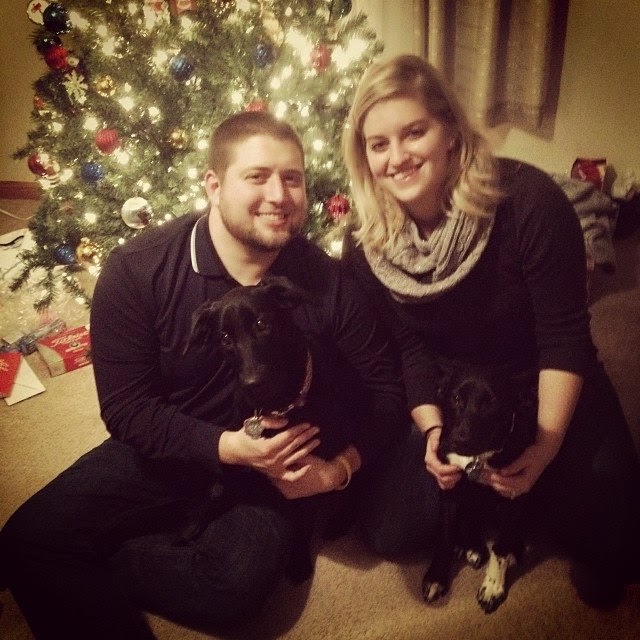
Me, my wife Kayla, I am holding Ellie, she is holding Hannah.
Sunday, July 9, 2017
Saturday, February 20, 2016
Update
So I haven't posted an update from my December 2015 doctors visit.
We are still good so far, nothing has grown. I do not have to go back for 6 months, so around May-June I will be heading back to Pittsburgh for another CAT Scan and another doctors visit. Below is my test result:
We are still good so far, nothing has grown. I do not have to go back for 6 months, so around May-June I will be heading back to Pittsburgh for another CAT Scan and another doctors visit. Below is my test result:
EXAM DATE: 12/23/2015 1:12 PM
PROCEDURE: CT LOWER EXTREMITY WITHOUT CONTRAST RIGHT
CLINICAL INDICATION: Age: 26 years . Gender: Male.
Stated history: " BONE NEOPLASM" Additional history: None.,
COMPARISON: CT right knee 09/02/2015
TECHNIQUE:
Thin section noncontrast CT images were obtained of the right knee
with reconstructions in multiple planes.
FINDINGS:
There is redemonstration of innumerable ovoid osteochondral bodies
scattered about the right knee consistent with synovial
osteochondromatosis. Several have migrated anteriorly into the
anterior aspect of the intercondylar notch. Several project over the
popliteus tendon sheath as well as within a small Baker's cyst. There
are most conspicuous in number along the lateral and posterior
lateral joint line, but are diffusely scattered about the knee.
There is moderate lateral patellar tilt with moderate marginal
osteophytes in the patellofemoral compartment. There is a small knee
joint effusion. There are moderate marginal osteophytes in the medial
and lateral compartments, mild spurring of the tibial spines and
intercondylar notch.
There is generalized osteopenia. No intraosseous bone lesions are
identified. There is no evidence of acute fracture or traumatic
malalignment. There is mild nonspecific subcutaneous stranding in the
posterior aspect of the knee. No additional soft tissue masses are
identified.
Impression
IMPRESSION:
1. Similar number of osteochondral bodies in the setting of synovial
osteochondromatosis, some of which have migrated into the anterior
aspect of the notch. There are most prominent laterally and
posterolaterally, and some are positioned within a small Baker's cyst
and the popliteus tendon sheath.
In other news, I started a new job at a company called EMKA at the beginning of February.
Also, if you like salsa, be sure to check out my business!
Pre-order the salsa: https://b-mys-homemade-famous-salsa-100-all-natural-salsa.backerkit.com/hosted_preorders
https://www.facebook.com/BMYsHomemade/
http://www.bmyshomemade.com/
Until Next Time,
Brian
Tuesday, September 22, 2015
1 Year Later
So it has been a year since I had my last surgery on September 22, 2014. So far it is still gone for the most part, there is still a piece lingering on the outer part of my leg, as shown in an X-Ray in May and a CAT scan I had earlier in September. So I have to go back to the doctors once again in December and get another CAT scan to make sure that it is not getting any bigger. Overall, I feel great, I did not even notice that there was something in my leg, and in my opinion, as long as it is not bother me, I would rather try to avoid having surgery again. I just need this tendentious to go away, but I have been working out with a trainer and I have been feeling the best I have in over 3 years. I also believe that my leg may never get straight again, but between that and tendentious, it is way better than having surgery constantly.
Here is the results from the most recent CAT scan:
Here is the results from the most recent CAT scan:
CT LWR EXTR WITHOUT CONTRAST RIGHT - Details
Narrative
CLINICAL HISTORY:
26-year-old male with osteochondral bodies seen on previous CR
imaging at lateral joint line and 04/15/2015.
TECHNIQUE:
Axial CT images of the patient's knee were obtained at 2.5 mm
intervals from the distal third of the femur through the proximal
tibia and fibula, examined on soft tissue and bone algorithm and used
as a basis for coronal and sagittal reformat images.
Comparison:
Previous CR imaging from 04/15/2015.
FINDINGS:
Multiple punctate areas of ossification are seen at the posterior
lateral aspect of the patient's knee joint prior CR imaging was
thought to represent multiple osteochondral bodies. There does not
appear to be an appreciable increase in the bodies compared to the CR
imaging from April of this year.
In addition, there is patchy osteopenia of the distal femur and tibia
which may be due to disuse or hyperemia.
There is mild narrowing of the medial compartment of the knee with
minimal osteophyte formation, and a similar degree of mild narrowing
and osteophyte formation of the lateral compartment and
patellofemoral compartment.
A small knee effusion is present. Extensor mechanism appears
unremarkable.
The coronal and sagittal reformatted images confirm the relationship
of the multiple osteochondral bodies at the posterior lateral aspect
of the knee with respect to the femur and tibia.
Impression
IMPRESSION:
No significant increase in multiple osteochondral bodies compared to
serial imaging from 05/15/2015.
No significant increase in multiple osteochondral bodies compared to
serial imaging from 05/15/2015.
So overall, it seems to be good news to me!
Sunday, May 31, 2015
It is still gone
Thought that I would give an update since it has been a while.
Went to the doctors in April and it is still clear, no disease. I go back again in Septemeber, and if it is still clear, I will start making yearly check-ups. Before I go back again, I have to get another MRI just to make sure it has no grown back.
Still really no stiffness, I have been going to physical therapy again to try and get rid of the tendonitis that I had in my knee, also to get rid of the tightness, which is a knot, in the back of my leg.
I am hoping to try and get some mountain biking in this year at some point.
Since my Kickstarter was successful, I now have a website for the business if you want to check it out!
www.bmyshomemade.com
Until next time,
Brian
Wednesday, January 14, 2015
And it is gone!!
Well today I drove to Pittsburgh for a check up, got X-Rays done, the doctor checked it came in and said well I wish your mom and wife were here to hear good news for once! According to the X-Rays, there is nothing left!! Praise God that it is gone!! So I go back in for another check up in 3 months to get it looked at again, assuming it stays away I will be good to go I think!
Now it is on to working out again like normal, I started up doing Shawn T's T25 and it is kicking my butt!! Then hopefully doing some mountain biking here in the spring!
I also have started up a Kickstarter to try and take my salsa making into a business! So if you wouldn't mind backing I would appreciate it, if you don't want to back, at least give my page a like!
https://www.kickstarter.com/projects/bmy/b-mys-homemade-salsa-100-all-natural-organic-salsa
https://www.facebook.com/BMYsHomemade
Sunday, December 7, 2014
It has been a while since the last update...
So since it has been almost 2 months since my last update, I have been doing great!! All I can say is that this is the best my knee has felt in 2 years!! Got the stitches out a few months ago, finished physical therapy before Thanksgiving, and started a new job this past Monday. I can actually squat now without much pain, still doing stretching on my own. I go back to the doctors here soon, which I am changing until early next year because of the new job. I still have some spots where I do not have feeling in my right knee. I will actually be surprised if it has grown back because it does not hurt at all, the tendonitis has seemed to have gone away for the most part and the stitches look great!
No need for pictures because there is nothing to see except for a scar. I can't wait until it is not winter anymore so I can get back to mountain biking.
Hopefully I won't wait for 2 months to update again.
No need for pictures because there is nothing to see except for a scar. I can't wait until it is not winter anymore so I can get back to mountain biking.
Hopefully I won't wait for 2 months to update again.
Saturday, October 4, 2014
2 Weeks after surgery
So it will be 2 weeks since my latest surgery on Monday. Just a quick update, I am still sore, I can feel the stitches pulling pretty good so I am sure they are ready to come out, which I will be getting that done on Wednesday. Some of them are already falling out on their own.
I am able to walk without crutches, I am still using an immobilizer when I walk up and down stairs and when I am outside. Mostly hurting in the front incision. In Physical Therapy they are moving the scar tissue around in my knee, I can tell that because when they are rubbing it, it is burning, they said that it is normal when the scar tissue is breaking up.
So here is a few pictures of the stitches of my knee from right after surgery.
Subscribe to:
Posts (Atom)